TRANSDIFFERENTIATION, DEDIFFERENTIATION, CELLULAR PLASTICITY, DYSPLASIA AND METAPLASIA: IS THE SPIKE PROTEIN CHANGING CELLULAR IDENTITY? COULD CARVACROL AND THYMOL BE POWERFUL THERAPEUTICS?
Perhaps the reason so much goes so wrong in so many systems is that cells are being reprogrammed
FROM
TO
Those who have followed my work for a long time know that I have been seeking a unifying mechanism for the pathology of COVID-19 and the Spike Protein. The more I learn, the more an image comes into focus. Recently, that focus has been on what could possibly make SO MUCH go SO WRONG? We have looked at fibrosis, senescence, brainstem damage, microvascular damage, neurological damage and the list seems to be endless and constantly expanding – and that’s the point! CONSTANTLY EXPANDING!
What I realized this evening, and have hinted at in a recent post, is that I now believe the major mechanism behind the pathology of COVID-19 and its Spike Protein is CELLULAR PLASTICITY. I believe that not only the Spike Protein, but perhaps the N and E proteins as well, are causing cells to CHANGE WHAT THEY ARE. This is being done through the processes of Transdifferentiation, Dedifferentiation, Dysplasia, Metaplasia and Cellular Plasticity itself.
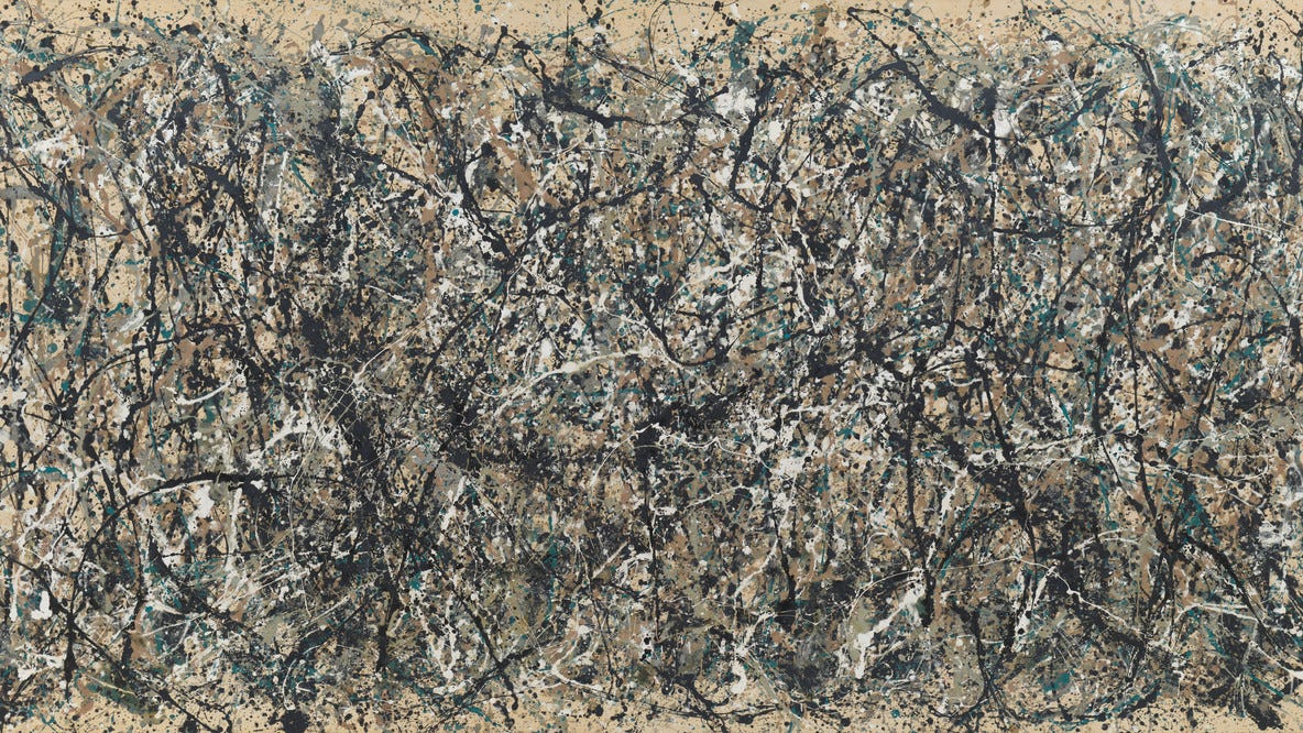
If you noticed, I began the post with a classical masterpiece of art: A Rembrandt. All highly structured and orderly. I won’t debate aesthetics, but to the average eye, the Pollock represents disorder and chaos. Imagine each molecule of paint in the Rembrandt being transformed into the Pollock. It is still PAINT but it is not the PAINT it began as. Such is what I now believe the Virus and its Proteins are doing.
THE EVIDENCE
In lungs, COVID-19 is affecting the differentiation of alveolar cells. This is also a hallmark of Idiopathic Pulmonary Fibrosis.
Recent single-cell RNA sequencing studies in acute alveolar injury mouse models have identified unique AT2 to AT1 transitional alveolar epithelial cell types after alveolar damage. These cells are defined variably as a Krt8+ alveolar differentiation intermediate (ADI), damage-associated transient progenitor (DATP), or pre-AT1 transitional state cell (PATS) (ADI/DATP/PATS hereafter). Incomplete transition from AT2 to AT1 cells, with an accumulation of ADI/DATP/PATS cells, has also been identified in human idiopathic PF (IPF) and in COVID-19 postmortem lungs, suggesting a common dysfunction in prolonged epithelial repair/disrepair.
SARS-CoV-2 infection produces chronic pulmonary epithelial and immune cell dysfunction with fibrosis in mice
https://www.science.org/doi/10.1126/scitranslmed.abo5070
In the brain, COVID is affecting the differentiation/plasticity of oligodendroglia lineage cells, resulting in loss of myelin plasticity.
Mild respiratory COVID impairs myelinating oligodendrocytes
We next explored the effects of mild respiratory COVID on oligodendroglial lineage cells. Reactive microglia/macrophages can alternatively promote (Miron et al., 2013) or impair (Gibson et al., 2019) oligodendrogenesis, depending on the precise microglial cell state. In disease states such as CRCI, reactive microglia cause a dysregulation of oligodendroglial lineage cells (Gibson et al., 2019) and loss of myelin plasticity (Geraghty et al., 2019).
Mild respiratory COVID can cause multi-lineage neural cell and myelin dysregulation
https://www.sciencedirect.com/science/article/pii/S0092867422007139#bib15
In the kidney, COVID affects the differentiation and plasticity of tubule cells.
After data processing and quality control, we obtained 20,165 single-cell transcriptomes. The most prominent finding in the snRNAseq analyses was in the proximal tubule (PT) compartment. We defined two cell populations corresponding to mature and undifferentiated PT cells, connected by two cell state transitions (Figure 1). Undifferentiated PT cells display an injured pattern characterized by metabolic impairment, reduction of the tubule transport function, and expression of injury markers confirmed in immunochemistry. We found that tubule repair follows two converging patterns involving the plasticity of mature tubule cells and the expansion and differentiation of progenitor-like cells. Tubule repair by cell plasticity displayed substantial similarities among mice and men and determined the transient expansion of undifferentiated tubule cells with altered functional and metabolic properties. Progenitorlike cells marked by PROM1 proliferated in response to injury and followed a differentiation process characterized by the sequential activation of the WNT, NOTCH, and HIPPO signaling pathways.
Single cell profiling in COVID-19 associated acute kidney injury reveals patterns of tubule injury and repair in human
https://pesquisa.bvsalud.org/global-literature-on-novel-coronavirus-2019-ncov/resource/pt/covidwho-1623121
In the pancreas, COVID is inducing the transdifferentiation of Beta-Cells. This may very well be why we are seeing de novo Diabetes post COVID.
Q&A with Shuibing Chen, PhD, Professor, Weill Cornell Medicine
What is your presentation about?
SARS-CoV-2 viral antigen is detected in beta cells of autopsies of COVID-19 subjects.
SARS-CoV-2-induced beta cell transdifferentiation is mediated by eIF2 pathway.
Trans-ISRIB reverses SARS-CoV-2 infection-induced beta cell transdifferentiation.
Proinflammatory macrophages are activated in SARS-CoV-2 infected human islets, as well as autopsies of COVID-19 subjects.
SARS-CoV-2 Infection Induces Beta-Cell Transdifferentiation
https://www.adameetingnews.org/live-updates/article/presenter-profiles/presenter-profiles-sars-cov-2-infection-induces-beta-cell-transdifferentiation/
In vasculature, COVID causes vascular cell dysplasia, endothelial dysfunction and a procoagulant state.
H-Hcy is associated with the development of atherosclerotic vascular disease including arterial hypertension and congestive heart failure. The underlying mechanisms are nitric oxide (NO) antagonism, production of reactive oxygen species (ROS), pro-thromboplastic activity, loss of blood vessel vasorelaxation and alterations in the elastin/collagen ratio. Further, there is a correlation between H-Hcy and arterial hypertension that especially applies to males as androgen hormones, in contrast to estrogen, increase ACE and angiotensin-receptor-1 (AT1R) activity. Altogether, these changes result in vascular cell dysplasia, endothelial dysfunction and a procoagulant state.
While these processes are slow and develop over years, acute H-Hcy activates a pro-inflammatory cascade through upregulation of the nuclear transcription factor (NF-kB) in neutrophils and macrophages, which release an ample amount of ROS potentiating oxidative stress.
Life-threatening course in coronavirus disease 2019 (COVID-19): Is there a link to methylenetetrahydrofolic acid reductase (MTHFR) polymorphism and hyperhomocysteinemia?
https://www.ncbi.nlm.nih.gov/pmc/articles/PMC7467063/
These dedifferentiation effects may also be a factor in the recent massive rise in new and aggressive cancers.
Researchers at the Ludwig Institute for Cancer Research discovered a mechanism that methodically reverts—or de-differentiates—cancer cells of a benign subtype of pancreatic tumors into a progenitor (immature) state of cellular development that proceeds to spawn highly aggressive tumors capable of metastasis to the liver and lymph nodes.
Research Shows Pancreatic Cancer Cells Reverse to Advance Malignancy
https://journals.lww.com/oncology-times/Fulltext/2021/07050/Research_Shows_Pancreatic_Cancer_Cells_Reverse_to.12.aspx
This mechanism may also be involved in some of the sudden deaths – as cardiac tissue is replaced by fatty or fibrofatty tissues.
Arrhythmogenic right ventricular dysplasia / cardiomyopathy (ARVD/C) is a rare familial disorder that may cause ventricular tachycardia and sudden cardiac death in young, apparently healthy individuals. The clinical hallmark of the disease is ventricular arrhythmias, arising predominantly from the right ventricle. The pathological hallmark of the disease is fibrofatty replacement of right ventricular myocardium.
Arrhythmogenic Right Ventricular Dysplasia/Cardiomyopathy (ARVD/C)
https://www.hopkinsmedicine.org/health/conditions-and-diseases/arrhythmogenic-right-ventricular-dysplasia--cardiomyopathy-arvdc
We may have a powerful therapeutic against this mechanism as Carvacrol and Thymol are protective of DNA changes.
At concentrations = IC20-40, the compounds studied did not induce DNA strand breaks either in human cells HepG2 or in cells Caco-2. Incubation of HepG2 and Caco- 2 cells in the presence of the whole scale of concentrations of carvacrol or thymol led in both cases to a significant protection of the cells studied toward DNA strand breaks induced by a potent oxidant hydrogen peroxide.
DNA-protective effects of two components of essential plant oils carvacrol and thymol on mammalian cells cultured in vitro
https://pubmed.ncbi.nlm.nih.gov/17319782/
Indeed, they have been thought to be effective against COVID.
Carvacrol is one of the monoterpene phenol with abundant presence in essential oils of many aromatic plants, including thyme and oregano. It is being used as food flavoring, additive, and preservatives. Carvacrol is also used as a fragrance in cosmetic products. A number of research studies have shown biological actions of carvacrol with its therapeutic potential is of clinical significance. The in vitro and in vivo studies have shown multiple pharmacological properties such as anticancer, anti-fungal, anti-bacterial, anti-oxidant, anti-inflammatory, vasorelaxant, hepatoprotective, and spasmolytic. This review highlights the various biological and pharmacological properties of carvacrol within the scope of COVID-19.
Carvacrol, a Plant Metabolite Targeting Viral Protease (Mpro) and ACE2 in Host Cells Can Be a Possible Candidate for COVID-19
https://www.frontiersin.org/articles/10.3389/fpls.2020.601335/full


Oregano has been used throughout Thyme as a great remedy...
Thank you Walter. Your strength and drive are an inspiration.