The Spike Protein and the Golgi Apparatus: A Mechanism for Induced Autoimmunity
The proteopathic Spike Protein fragments the Golgi Apparatus, likely inducing autoimmune disease.
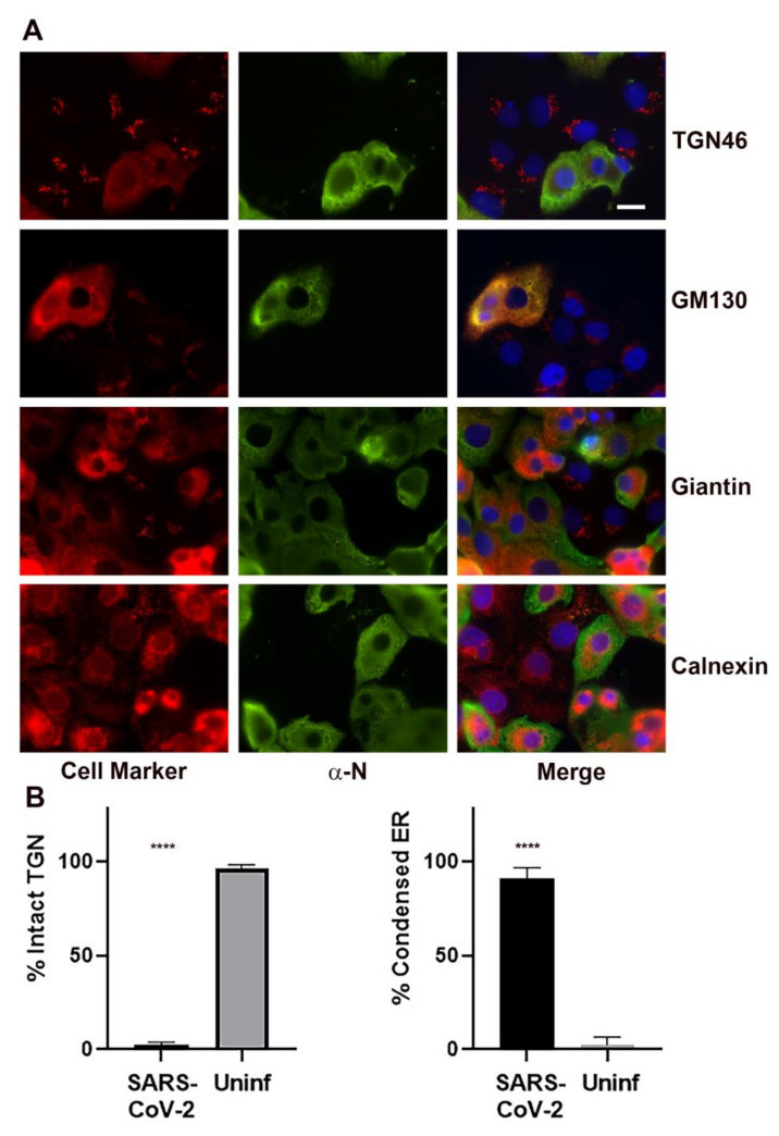
(B) Quantitation of the TGN46 observed in cells infected with SARS-CoV-2 vs. uninfected control cells. TGN46 localization was determined for 315 SARS-CoV-2 infected cells in three biological replicates and 522 uninfected control cells in five biological replicates. For ER condensation, 125 SARS-CoV-2 infected cells and 106 uninfected control cells in five biological replicates were analyzed. Shown is the mean ± the S.E.M. Statistics were performed using One-way ANOVA. Significant differences relative to the uninfected control are indicated (**** p < 0.0001).
One of the largest conundrums of post-SARS-CoV-2 infection and Spike Protein exposure is the observed incidence of autoimmune disease. In particular, the autoimmune response bears very close resemblance to Sjogren’s Syndrome and Lupus. Why does this happen? The answer may be found in the Spike Protein’s (and other viral proteins) ability to fragment and destroy the Golgi Apparatus.
A remarkable finding 24h post SARS-CoV-2 infection was the following:
Quantitation of the frequency of Golgi dispersal revealed that by 24 h post-infection, the Golgi apparatus was intact in 2.2 ± 0.8% of the infected cells compared to 96.6 ± 0.8% in the uninfected cells.
Knowing that the Golgi Apparatus is fragmented and destroyed is one thing. Knowing what causes it is of even greater importance. Indeed, the Spike Protein does cause the fragmentation and destruction of the Golgi Apparatus.
In an effort to identify specific viral proteins that might be responsible for disruption of the Golgi apparatus, we transfected Vero cells with an expression library of SARS-CoV-2 proteins. Interestingly, five of the expressed proteins, M, S, E, Orf6, and nsp3, caused dispersal of the Golgi apparatus.
Disruption of the Golgi Apparatus and Contribution of the Endoplasmic Reticulum to the SARS-CoV-2 Replication Complex
https://www.ncbi.nlm.nih.gov/pmc/articles/PMC8473243/
The fragmentation of the Golgi Apparatus can have very serious consequences. What I find compelling is that both Sjogren’s Syndrome and Systemic Lupus Erythematosus can be induced by Golgi Apparatus fragmentation.
Anti-Golgi complex autoantibodies are found primarily in patients with Sjögren's syndrome and systemic lupus erythematosus, although they are not restricted to these diseases.
(…)These data point to a crucial role for fragments of autoantigens in the generation of autoantibody responses. In the present study, several Golgi autoantigens were detected cleaved into distinctive fragments during apoptosis and necrosis. It has been speculated that these modified forms of autoantigens may have enhanced immunogenicity through exposure of immunocryptic epitopes that are not generated during antigen processing. These epitopes could trigger autoimmune responses if presented to the immune system under proinflammatory conditions, and they may be recognized as surface structures on cytoplasmic organelles that are released to the immune system in aberrant disease states.
Autoantibody responses could be amplified and maintained on repeated stimulation if the exposure of intracellular antigens to the immune system is associated with defective clearance of apoptotic cells, prolonged necrosis (primary or secondary), T-cell cytotoxicity associated with chronic infection, or even antigen mutation or overexpression.
Fragmentation of Golgi complex and Golgi autoantigens during apoptosis and necrosis
https://www.ncbi.nlm.nih.gov/pmc/articles/PMC125295/
Indeed, we are observing something akin to Sjogren’s and Lupus post COVID (Spike Protein) exposure.
Sjögren’s Disease (SjD) is a chronic and systemic autoimmune disease characterized by lymphocytic infiltration and the development of dry eyes and dry mouth resulting from the secretory dysfunction of the exocrine glands. SARS-CoV-2 may trigger the development or progression of autoimmune diseases, as evidenced by increased autoantibodies in patients and the presentation of cardinal symptoms of SjD.
Evidence of a Sjögren’s disease-like phenotype following COVID-19
https://www.ncbi.nlm.nih.gov/pmc/articles/PMC9628191/
There have been some reported cases of autoimmune disease development after COVID-19 infection. We present a patient with a history of COVID-19 infection one month prior who presented with non-specific symptoms including fatigue, malaise, bilateral lower extremity swelling and shortness of breath. His laboratory evaluation and physical exam showed him to be in acute renal failure. Further workup and kidney biopsy results confirmed systemic lupus erythematosus (SLE).
New onset systemic lupus erythematosus after COVID-19 infection: a case report
https://www.ncbi.nlm.nih.gov/pmc/articles/PMC9010314/
The fact that the autoimmune response is “Lupus-like” I believe further supports this hypothesis.
Researchers are still trying to determine whether long Covid is itself an autoimmune condition or if Covid can trigger a secondary autoimmune disease like lupus or rheumatoid arthritis for which some patients haven't been properly diagnosed.
Autoimmune diseases can be hard to detect, since many have loose or overlapping definitions. There's no single test to diagnose lupus or rheumatoid arthritis.
Mounting evidence shows autoimmune responses play a significant role in long Covid
https://www.nbcnews.com/health/health-news/long-covid-autoimmune-response-rcna48917
I have previously written that the Spike Protein executes a “Sherman’s March through Georgia” during its cellular rampage. The devastation is secondary, I believe, or perhaps A CONSEQUENCE OF its ability to install itself as a systemic proteopathic seed.


At the very outset the creation of an autoantigen seemed a very, very bad idea indeed. Thank you for helping flesh out the details where the devil resides.
They knew what they were doing Walter . The discussion with Kevin McCairn was great . Thanks Walter!