AFTER DESTROYING THE ENDOTHELIUM, THE SPIKE PROTEIN, IN ADDITION TO INDUCING FIBROSIS, THEN TURNS THE VASCULATURE TO “IRON AND STONE”
THE SPIKE PROTEIN IS INDUCING THE CALCIFICATION OF AND HEMOSIDERIN DEPOSITION INTO THE VASCULATURE: THE KEY TO THE WHITE CLOTS
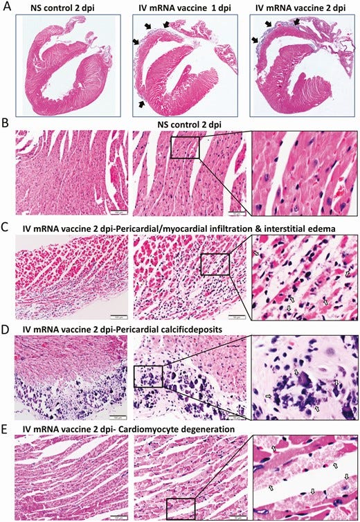
I read a paper from a group in Hong Kong that had injected a Spike Protein Accelerant into mice. With the surprising result that within two days THE HEART WAS SIGNIFICANTLY CALCIFIED. So, I decided to look into this a little deeper.
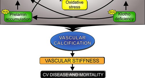
It turns out that Cellular Senescence, Decreased Autophagy and most importantly EXTRACELLULAR VESICLES contribute to Vascular Calcification in Aging. These are exactly the mechanisms that the Spike Protein uses and/or induces.
This may be the underlying mechanism to all the Post COVID and Post Spike Protein pathologies, including Long COVID. The vasculature may be aged fifty years by the Spike Protein.
More disturbingly, in a recent study, it was found that the CaMKII-like domains of the S protein mediated the fusion of virus and infected cells, and it involved calcium ions in this process. In membrane fusion, calcium ions near the interface between S protein and cell membrane quickly accumulated, which produced abnormal calcium ion currents. Besides that, abnormal flow of calcium ions caused depolarization of cells near the infected cell, prompting many calcium ions to flush into nearby cells. Finally, the calcium ion demand in serum accelerated in a short time, thus facilitating the release of calcium in the body. If the SARS-CoV-2 virus infection area was large, it encouraged the abnormality of serum calcium levels and disrupted the body’s calcium homeostasis.
Concluding that membrane fusion is increasing the risk of coagulation and VASCULAR CALCIFICATION.
On top of all of this, results of seven autopsies were as follows: Seven deceased COVID-19 patients underwent MIA with brain MR and CT images, six of them with tissue sampling. Imaging findings included infarcts, punctate brain hemorrhagic foci, subarachnoid hemorrhage and signal abnormalities in the splenium, basal ganglia, white matter, hippocampi and posterior cortico-subcortical. Punctate brain hemorrhage was the most common finding (three out of seven cases). Brain histological analysis revealed reactive gliosis, congestion, cortical neuron eosinophilic degeneration and axonal disruption in all six cases. Other findings included edema (5 cases), discrete perivascular hemorrhages (5), cerebral small vessel disease (3), PERIVASCULAR HEMOSIDERIN DEPOSITS (3), Alzheimer type II glia (3), abundant corpora amylacea (3), ischemic foci (1), periventricular encephalitis foci (1), periventricular vascular ectasia (1) and fibrin thrombi (1). SARS-CoV-2 RNA was detected with RT-PCR in 5 out of 5 and IHC in 6 out 6 patients (100%).
In a patient that survived, a repeat MRI of the brain without contrast agent enhancement showed residual T2 hyperintensities and hemosiderin deposition in the medial thalami; T2 hyperintensities significantly improved and HEMOSIDERIN DEPOSITION REMAINED UNCHANGED.
This would indicate that subsequent exposures to the Spike Protein would incrementally increase the amount of iron deposited into the vasculature. And how do we know it is Spike? The following is very telling: SARS-CoV-2 spike protein was also detected in cytoplasm of cutaneous dermal vessels and eccrine cells on immune-histochemistry (IHC) of a 35-years-old man presenting with acral purpuric macules although his RT-PCR was negative. As the lesions evolve, the color changes to copper red and violaceous due to associated red cell extravasation and then orange-brown as a result of HEMOSIDERIN DEPOSITION.
This Perivascular Hemosiderin Deposition also occurs in MS! Where it causes a VASCULITIS! Hemosiderin deposition was common in the substantia nigra and other pigmented nuclei in all cases. It is concluded that the cerebral vein wall in multiple sclerosis is subject to chronic inflammatory damage, which promotes haemorrhage and increased permeability, and constitutes a form of vasculitis.
And the white clots? Calcium!
This may be the answer to them as well. The dimeric FXIII-A2, a pro-transglutaminase is the catalytic part of the heterotetrameric coagulation FXIII-A2B2 complex that upon activation by calcium binding/thrombin cleavage covalently cross-links preformed fibrin clots protecting them from premature fibrinolysis. Note, that in COVID, a comparison between healthy plasma and acute COVID-19 solubilized clots also showed a significant increase in coagulation factor XIII A chain, VWF Complement component C7 and CRP.
Yes, it is this bad. Please stop all the “all fear, no solutions” mantras against me. We cannot even begin to find solutions until we know what it is actually doing. Eventually we have to reach the bottom of this pit. Let’s just hope that isn’t the discovery that infection or transfection with the Spike Protein of SARS-CoV-2 ultimately results in 100% mortality. Believe me. I do not want to find that.
You MUST remember. HIV starts with a “mild flu-like illness.” Stop calling COVD a damn cold. And we ABSOLUTELY MUST stop forcing Spike Protein Accelerants on the Known Universe!
Referenced/Related Papers
https://pubmed.ncbi.nlm.nih.gov/3346691/
https://www.ncbi.nlm.nih.gov/pmc/articles/PMC8374125/
https://pubs.rsna.org/doi/full/10.1148/radiol.2020203132
https://onlinelibrary.wiley.com/doi/10.1111/dth.14951
https://insightsimaging.springeropen.com/articles/10.1186/s13244-021-01144-w


Walter, forgive my ignorance, as I have no bio training and have been reading your sub-stack enthusiastically for the past couple months, trying to learn and absorb as best I can. I have noticed a marked refocus on the actual C19 virus in your focus of concern, as opposed to the gene therapy "vaccinations." In fact I've seen the same attention paid to the virus, as of late, with Igor Chudov as well (the two of you are my fave bio-investigators).
What I'm hearing from both of you on the virus is starting to really concern me, especially since my wife, both kids, and I had Omicron back in January. We are all vaxx-free, and the immediate external effects at the time were relatively mild. Stupidly, I only had enough Ivermectin on hand at the time to dose my wife and I, and not my children...we did so only because the higher risk of our age groups over our children (at least as we understood at the time). I'm now regretting that decision and thinking (in light of all you have revealed lately) that my kids would have been better off getting the IVM.
All this time I thought, so long as we didn't get the vaxx, and could fight of the infection if we ever got it, then we'd be set with good immunity and no worries about the side effects of the vaxx. In other words my fear was always about the vaxx, and not so much the virus since we're a quite healthy family. Now, from the tone of what I'm hearing, the virus may even be the bigger danger long term.
I know you've mentioned the vaccine as a spike accelerator, so it would at least partially calm my nerves if I knew we'd be way worse off if we were vaccinated. It's just that right now, I'm thinking I was looking in the wrong direction the whole time. Are you able to at least comment on where you are striking the danger balance of probabilities between the virus itself and the vaccine. Is it the virus after-all that is the real danger, notwithstanding the additional complications of ADE and the like?
Thanks for all your hard work, Walter! You are an amazing person.
Kaz
This looks promising: https://patents.google.com/patent/US8309081B2/en
Primarily a combination of chelator and proteolytic enzyme, along with things that strengthen blood vessels and recruit white blood cells.
Here's a commercial product that uses arnica, IP6, and bromelain: https://www.scarguard.com/products/bruise-fader