

The Spike Protein Parallels Asbestos: Malignant Biphasic Mesothelioma Post mRNA
As I predicted in 2021, the Spike Protein mimics the effects of Asbestos in the body.

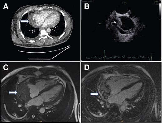
(A) Contrast computed tomography thorax (using Omnipaque 350 contrast agent) showing a filling defect in the right atrium’s lateral wall. (B) This was also seen in the transoesophageal echo as seen in the image on the right. (C) Four-chamber balanced SFPP Cine sequence revealed a large mass (6 × 3 cm) with irregular margins which appears to originate from the pericardium and compressing the right atrium. (D) Four-chamber delayed gadolinium enhancement sequence showed heterogeneous enhancement of the mass and no myocardial late gadolinium enhancement.
Four years ago, I realized that the Spike Protein behaves in precisely the same way as Asbestos does in the body.
In fact, I believe the Spike Protein causes an “accelerated asbestosis.” I have substituted Spike Protein for Asbestos in the following:
When Spike Proteins reach the alveoli in the lung, where oxygen is transferred into the blood, the foreign bodies (Spike Proteins) cause the activation of the lung’s local immune system and provoke an inflammatory reaction. This inflammatory reaction can be described as chronic rather than acute, slow ongoing activation of the immune system in an attempt to eliminate the Spike Proteins. Macrophages phagocytose the Spike Proteins and stimulate fibroblasts to deposit connective tissue. Due to the natural resistance of Spike Proteins to digestion, the macrophage dies off, releasing cytokines and attracting further lung macrophages and fibroblastic cells to lay down fibrous tissue, which eventually forms a fibrous mass. The result is interstitial fibrosis. The fibrotic scar tissue causes alveolar walls to thicken, which reduces elasticity and gas diffusion, reducing oxygen transfer to blood as well as the removing of carbon dioxide.
The Spike Protein and Parallels to Asbestos
https://wmcresearch.org/the-spike-protein-and-parallels-to-asbestos/
Today, unfortunately, we have evidence that this is, in fact, the case. A just published case series report details how, four weeks after his first Pfizer–BioNTech COVID-19 vaccine, a 49-year-old male was diagnosed with malignant biphasic mesothelioma. He died four months later.
In Case 1, a 49-year-old Black African male presented with chest pain and breathlessness after a COVID-19 vaccine. Initially treated for pericarditis, he returned with worsening symptoms. Echocardiography revealed pericardial effusion and cardiac tamponade. Imaging confirmed a right atrial mass diagnosed as malignant biphasic mesothelioma. He died 4 months after diagnosis. In Case 2, a 43-year-old Caucasian male developed breathlessness and fever post-COVID-19 vaccine. Imaging identified a large posterior pericardial mass, later diagnosed as synovial sarcoma. Chemotherapy yielded minor tumour reduction, but he succumbed to his illness, spending his final days in a hospice.
Heart-breaking tumours: a case series of malignant pericardial effusion
https://academic.oup.com/ehjcr/article/9/3/ytaf009/7960074
Why is this so alarming? Because Mesothelioma is almost exclusively caused by Asbestos - a degradation-resistant fiber.
Mesothelioma is almost always caused by exposure to asbestos, a group of minerals made of microscopic fibres that used to be widely used in construction.
These tiny fibres can easily get in the lungs, where they get stuck, damaging the lungs over time.
It usually takes a while for this to cause any obvious problems, with mesothelioma typically developing more than 20 years after exposure to asbestos.
Mesothelioma
https://www.nhs.uk/conditions/mesothelioma/
Of course, Mesothelioma it is normally related to the lungs – because you inhale it. However, it should come as no surprise that we now find it in the heart, because mRNA goes... everywhere.
We also gain further understanding of the mechanisms involved. This may be useful for determining systemic effects and developing treatments.
Asbestos may cause Mesothelioma via Ferroptosis.
The deposition of asbestos fibers in tissues offers a surface where iron-reach macromolecular aggregates (asbestos bodies) favoring the development of chronic inflammation (15). Moreover, it has been shown that asbestos-activated macrophages release ROS that, in turn, may induce DNA damage indirectly via formation of 8-hydroxy-2’-deoxyguanosine (8-OHdG) adducts (16). Recent data on iron-catalyzed ROS production suggests that ferroptosis, a non-apoptotic, iron-dependent cell death, may be involved in asbestos-related carcinogenesis (17). Moreover, a role in asbestos-induced carcinogenesis has been postulated for hepatocyte growth factor (HGF), via activation of the PI3K/MEK5/Fra-1 axis (18).
On the other hand, the exposure to asbestos fibers causes death of human mesothelial cell (HM), a cell type particularly susceptible to fiber cytotoxicity that was initially ascribed to apoptosis (19). Afterwards, asbestos pathogenesis was clearly associated with tumor necrosis factor-alpha (TNF-α), a mediator of inflammation (20).
How asbestos and other fibers cause mesothelioma
https://pmc.ncbi.nlm.nih.gov/articles/PMC7082251/
And, the Spike Protein is a master of Ferroptosis.
Viral RNA and spike protein were detected in SAN cells in the hearts of infected hamsters. We established an efficient strategy to derive from hESCs functional human SAN-like pacemaker cells, which express pacemaker markers and display SAN-like action potentials. Furthermore, SARS-CoV-2 infection causes dysfunction of human SAN-like pacemaker cells and induces ferroptosis.
SARS-CoV-2 Infection Induces Ferroptosis of Sinoatrial Node Pacemaker Cells
https://pmc.ncbi.nlm.nih.gov/articles/PMC8963443/
Coronavirus disease 2019 (COVID-19) patients show lipid metabolic alterations, but the mechanism remains unknown. In this study, we aimed to investigate whether the Spike protein of severe acute respiratory syndrome coronavirus 2 (SARS-CoV-2) impairs lipid metabolism in host cells. We generated a Spike cell line in HEK293 using the pcDNA vector carrying the Spike gene expression cassette. A control cell line was generated using the empty pcDNA vector. Gene expression profiles related to lipid metabolic, autophagic, and ferroptotic pathways were investigated. Palmitic acid (PA)-overload was used to assess lipotoxicity-induced necrosis. As compared with controls, the Spike cells showed a significant increase in lipid depositions in cell membranes as well as dysregulation of expression of a panel of molecules involving lipid metabolism, autophagy, and ferroptosis. The Spike cells showed an upregulation of nuclear factor erythroid 2-related factor 2 (Nrf2), a multifunctional transcriptional factor, in response to PA. Furthermore, the Spike cells exhibited increased necrosis in response to PA-induced lipotoxicity compared to control cells in a time- and dose-dependent manner via ferroptosis, which could be attenuated by the Nrf2 inhibitor trigonelline. We conclude that the Spike protein impairs lipid metabolic and autophagic pathways in host cells, leading to increased susceptibility to lipotoxicity via ferroptosis which can be suppressed by a Nrf2 inhibitor. This data also suggests a central role of Nrf2 in Spike-induced lipid metabolic impairments.
The Spike Protein of SARS-CoV-2 Impairs Lipid Metabolism and Increases Susceptibility to Lipotoxicity: Implication for a Role of Nrf2
https://www.mdpi.com/2073-4409/11/12/1916
The most urgent matter is persistence of Spike. This is proven in Long COVID. Persistence of fibers that don’t degrade (Asbestos) is the reason for Mesothelioma and all things Asbestos. We must determine if this protein is hiding in viral reserves within the body, or if it is even being retrotranscribed. Again, the repeated introduction of Spike Protein into the body is beyond horrific. It is tantamount to putting our cells on the rack.
Thank you, as always, for your readership, dialog and support. You keep me going. And it is my honor and pleasure to seek, find and share with you.
I've been following Walter's posts intermittently for 2 years. Recently I think there is a good possibility that Marc Girardot and his Bolus Theory is correct regarding the causes of the blood clots and the damage to the endothelial system. If the needle is being accidently injected directly into veins in some proportion of people then the mRNA vaccine has access to all the cells that line the blood vessels. And then T cells detect those cells that are infected by mRNA vaccine and kill them. This could be the cause of all the blood clots. Does Walter address this theory in any way? It would be useful to gather up all Walter's postings and then use artificial intelligence to figure out things like this.
Marc Girardot and his Bolus Theory:
https://substack.com/@thebolustheory/p-152659010
My mother died in 1980 at 57 years old. She always smoked Kent cigarettes with their micronite filters. They were supposed to be safer because the filter would filter tars "mo bettah". Well, those filters were made from blue asbestos. https://csts.ua.edu/micronite/ https://en.wikipedia.org/wiki/Kent_(cigarette) Ad: https://www.youtube.com/watch?v=sKPUqKq_Bso