Friday Hope: Quercetin and Curcumin Inhibit Syncytia Formation
Quercetin and Curcumin may prevent and treat the fusion of cells, which may be driving sudden cardiac deaths and neurological issues due to the Spike Protein.
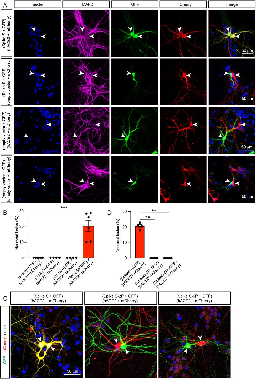
Expression of spike S and its receptor hACE2 induce fusion of murine neurons in culture.
a, Representative images of fused neurons (first row), or non-fused control neurons (other rows). Two populations of hippocampal neurons expressing a combination of two plasmids as indicated on the left (spike S and GFP, hACE2 and mCherry, empty vector and GFP, or empty vector and mCherry) were cultured together for 7 days (7 DIV). Immunocytochemistry for nuclei (blue), MAP2 (magenta), GFP (green) and mCherry (red). Neuronal fusion only occurred (first row) when one population of neurons was transfected with spike S and GFP, and the other with hACE2 and mCherry, as visualized by the presence of GFP and mCherry in the same neurons (yellow in the merge panel). b, Quantification of neuronal fusion as the percentage of neurons that fuse (yellow) when two neurons are in proximity ( ≤ 200 μm). c, Representative images of fused neurons (first panel), or non-fused neurons (second and third panels). Two populations of hippocampal neurons expressing a combination of two plasmids as indicated above the images (spike S and GFP, hACE2 and mCherry, spike S-2P and GFP, or spike S-6P and GFP) were cultured together for 7 days (7 DIV). Immunocytochemistry for nuclei (blue), MAP2 (magenta), GFP (green) and mCherry (red). Neuronal fusion only occurred (first panel) when the full-length WT spike S protein was transfected, as visualized by the presence of GFP and mCherry in the same neurons (yellow in the panel), and not when any of the non-fusogenic mutants (spike S-2P, spike S-6P) were used (second and third panels). d, Quantification of neuronal fusion as the percentage of neurons that fuse (yellow) when two neurons are in proximity ( ≤ 200 μm). Data in b and d were displayed as mean ± SEM, n > 200 neurons analyzed in 4-6 independent dishes from 2 dissections, one-way ANOVA Kruskal-Wallis test in e followed by Dunn’s post hoc test comparing all groups to the group without spike S or hACE2. **p <0.01, ***p <0.001.
If you have been reading my recent posts, you will know that I believe the fusogenicity of the Spike Protein of SARS-CoV-2 may be responsible for much of the sudden cardiac death and neurological issues being observed. Indeed, the Spike Protein causes fusion between neurons and glial cells.
Numerous enveloped viruses use specialized surface molecules called fusogens to enter host cells. During virus replication, these fusogens decorate the host cells membrane enabling them the ability to fuse with neighboring cells, forming syncytia that the viruses use to propagate while evading the immune system. Many of these viruses, including the severe acute respiratory syndrome coronavirus 2 (SARS-CoV-2), infect the brain and may cause serious neurological symptoms through mechanisms which remain poorly understood. Here we show that expression of either the SARS-CoV-2 spike (S) protein or p15 protein from the baboon orthoreovirus is sufficient to induce fusion between interconnected neurons, as well as between neurons and glial cells. This phenomenon is observed across species, from nematodes to mammals, including human embryonic stem cells-derived neurons and brain organoids. We show that fusion events are progressive, can occur between distant neurites, and lead to the formation of multicellular syncytia. Finally, we reveal that in addition to intracellular molecules, fusion events allow diffusion and movement of large organelles such as mitochondria between fused neurons.
What was very interesting about this study was that the Spike used in the vaccines did NOT cause cells to fuse, as it is in prefusion conformation, blocking its fusion capacity. Yet, they ONLY used hACE2. Also, Furin is able to cleave the spike, and it attaches to FAR MORE than just ACE2, I believe this needs to be further explored.
Unlike p15, the presence of the specific receptor was required to initiate cellular fusion, as expression of spike S or hACE2 alone did not generate any fusion events (Fig. 3a second and third rows, and b). To determine whether the fusion of neurons was caused by the fusogenic properties of spike S, we used two fusion-inactive versions of this protein, the spike S-2P and the spike S-6P (HexaPro, the universal vaccine Spike).
The SARS-CoV-2 spike (S) and the orthoreovirus p15 cause neuronal and glial fusion
https://www.biorxiv.org/content/10.1101/2021.09.01.458544v1.full
The ability to fuse cells does not appear to be limited to neurons and glial cells. Multinucleated cells attributed to the Spike have been found in the lungs and vascular walls of patients.
An additional pathology characteristic of COVID-19 disease was the presence of anomalous epithelial cells. These were characterized by abnormally large cytoplasm and, very commonly, by the presence of bi- of multi-nucleation. Presence of these dysmorphic cells was detected in the lungs of 20 patients (50%), including all 6 patients requiring IC, and occasional in additional 16 patients (39%). These cells were present in the alveolar spaces or jutted out into the neighboring, damaged vascular wall. In several instances, these cells clustered to form areas of squamous metaplasia.
Persistence of viral RNA, pneumocyte syncytia and thrombosis are hallmarks of advanced COVID-19 pathology
https://www.ncbi.nlm.nih.gov/pmc/articles/PMC7677597/
It should be noted that squamous metaplasia is a risk for cancer.
As we are discovering, the ability of the Spike to fuse cells also occurs in the heart, which is the basis for my concern that cardiomyocyte fusion is a major factor in the plethora of sudden cardiac deaths.
Transfection of hiPSC-CMs with CoV-2 S-mEm produced multinucleated giant cells (syncytia) displaying increased cellular capacitance (75±7 pF, n = 10 vs. 26±3 pF, n = 10; P<0.0001) consistent with increased cell size.
SARS-CoV-2 spike protein-mediated cardiomyocyte fusion may contribute to increased arrhythmic risk in COVID-19
https://journals.plos.org/plosone/article?id=10.1371/journal.pone.0282151
There is ample evidence proving that the Spike Protein is fusing cells together, potentially in every organ. Therefore, we must find a way to either treat or prevent the fusing of cells, to avoid potential serious harm and damage.
Once again, Quercetin and Curcumin come to the rescue. We find yet another way these wonder-molecules are able to assist us in our battle against this dangerous pathogen.
QUERCETIN
A study published in March of last year showed that Quercetin not only reduced syncytia formation, but also reduced spike protein expression. A double-benefit.
Findings
Spike transfection in HEK293-ACE2+ cells caused syncytia formation, cellular sloughing, and focal denudation of the cell monolayer; transfection in Vero E6 cells also caused syncytia formation. Spike expression upregulated potentially nephrotoxic genes (TNF-α, MCP-1, and ICAM1). Spike upregulated the cytoprotective gene HO-1 and relevant signaling pathways (p-Akt, p-STAT3, and p-p38). Quercetin, an HO-1 inducer, reduced syncytia formation and spike protein expression.
Interpretation
The major conclusions of the study are: 1) Spike protein expression in kidney cells provides a relevant model for the study of maladaptive and adaptive responses germane to AKI in COVID-19; 2) such spike protein expression upregulates HO-1; and 3) quercetin, an HO-1 inducer, may provide a clinically relevant/feasible protective strategy in AKI occurring in the setting of COVID-19.
The spike protein of SARS-CoV-2 induces heme oxygenase-1: Pathophysiologic implications
https://www.ncbi.nlm.nih.gov/pmc/articles/PMC8669938/
CURCUMIN
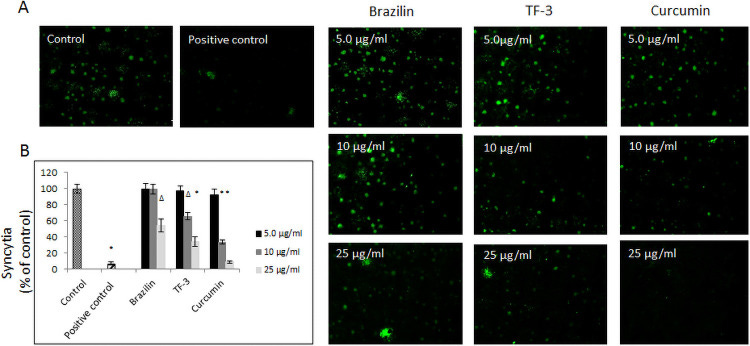
Effect of selected polyphenols on fusion to human ACE2 receptor overexpressing A549 cells.
(A) Cell-cell fusion of A549 cells expressing eGFP spike protein with A549 cells stably expressing human ACE2 receptor. The scale bar indicates 250 μm. (B) Quantitative analysis of formed syncytia. Experiments were done in triplicate and repeated three times. Data are presented as % of control ± SD; TF-3 –theaflavin-3,3’-digallate Δ p ≤ 0.01, * p ≤ 0.001. Control– 0.025% DMSO, positive control– 20 μg/ml anti-ACE2 antibody.
In a study from June 2021, it was found that, of tested polyphenols, Curcumin had the greatest effect in preventing the formation of syncytia.
Also, our further experiment, where A549 cells expressing SARS-CoV-2 spike protein pseudo-typed virions were pre-incubated with the tested polyphenols, and then layered for 4h on hCE2/A549, the cells showed a significantly decreased attachment. Incubation with brazilin at 25 μg/ml decreased the fusion by 40%, with TF-3 at 10–25 μg/ml by 40% to 70%, and with curcumin at the same concentrations, i.e., 10–25 μg/ml, by 70% to 95%. The results were consistent with previously obtained sets of data.
Phenolic compounds disrupt spike-mediated receptor-binding and entry of SARS-CoV-2 pseudo-virions
https://www.ncbi.nlm.nih.gov/pmc/articles/PMC8211150/
We need further studies to determine the best course of action in using compounds like Quercetin and Curcumin in treating Spike-related pathologies.
I would like to thank Joachim Gerlach and the team at vedicinals.com for their contribution to the research of this post. Vedicinals reached out to me as they discovered my treatment recommendations were very much in line with their own findings. They have a therapeutic which may prove to be a virtual protocol itself. It is called Vedicinals9.
https://www.vedicinals.com/


I know many people are already taking Vit C and NAC, and I don't vouch for the veracity of this article, I leave that to minds trained in these fields, I thought this article interesting.
The Quercetin Paradox-Toxicity
" This duality in quercetin—and its potential to be metabolized down one of two very different pathways—is known as the "quercetin paradox." Because quercetin can oxidize so easily, forming pro-oxidant, toxic metabolites, special care must be given to how quercetin-containing products are formulated. This is why quercetin should always be co-administered with vitamin C and NAC."
https://www.lifestylematrix.com/blog/post/the-quercetin-paradox-the-secret-to-preventing-toxic-quercetin-metabolites
Burdock Root (as dried or roasted(sweeter)) is a great source of quercitin. It helped me through a mystery illness late 2019. Burdock also has (among a 100 other things) inulin to help stabilise blood sugar which really helps with the appetite killing property of this 'mystery' disease.