Disturbing Evidence Supporting My 2021 Hypothesis that SARS-CoV-2 (Spike) Turns Us Into "Living Cartilage"
120dpi study in hamsters shows how SARS-CoV-2 dysregulates tissue repair and regeneration causing collagen deposition, fibrosis and cancer: Spike + Notch Activation
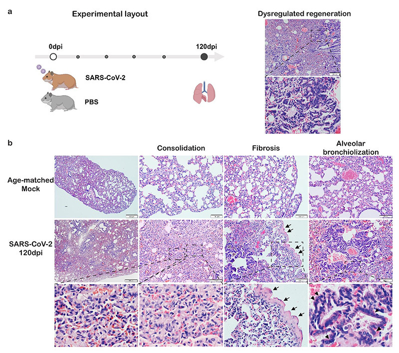
SARS-CoV-2 infection causes persistent abnormal foci of alveolar bronchiolization and fibrosis in hamster lungs. a. Experimental layout: 6-8 weeks old hamsters were intranasally inoculated with 103 PFU SARS-CoV-2 wild-type strain HK-13 or equal volume of PBS as mock controls. Lung tissues were collected at 120dpi. Dysregulated regeneration was observed in SARS-CoV-2 infected hamster lungs. Upper H&E image showing lung condensation with blood vessel congestion and multiple abnormal foci. Lower zoomed image showing abnormal foci of alveolar bronchiolizaiton. b. Representative H&E images showing pulmonary consolidation, fibrosis and alveolar bronchiolization in SARS-CoV-2 infected hamster lungs at 120dpi. Upper panel: Mock control lung showed normal structure and lung was not inflated. Middle panel: SARS-CoV-2 infected hamsters displayed whole lung condensation and multiple foci from proximal to distal lung. Bottom panel: Higher magnification and zoomed images showing alveolar collapse, alveolar consolidation, pleurae thickening and alveolar bronchiolization. Black open arrows indicated pleurae thickening. Black triangles indicated alveolar epithelial cell hyperplasia. Scale bar=500μm, 200μm, respectively.
I dedicate this post to my wonderful colleague Annelise Bocquet-Garçon.
First, do not be skeptical of the hamsters' presence. We need them here. There are currently no longitudinal studies of the mechanisms discussed here in human tissues.
In September of 2021 I made a finding which disturbed me greatly. I predicted that the Spike Protein, through signaling, would induce aberrant tissue repair and regeneration resulting in organs being turned into “cartilage.”
A MASS OF "LIVING" CARTILAGE?
https://x.com/Parsifaler/status/1440528972698177542
A paper published yesterday strongly supports this finding.
My initial finding was based on the Spike Protein activating Wnt5a signaling, this, in turn, activates NOTCH, which is responsible for dysregulated tissue regeneration and allows for the induction of cancer.
To investigate if the potentially Notch dysregulation induced bronchiolization caused by SARS-CoV-2 infection is possible risk factor for lung cancer, we conducted spatial transcriptomic experiment using hamster lung samples collected at 120dpi, comparing the transcriptomics profile of the histologically abnormal lung regions with that of the normal regions. Six tissue regions were identified and selected to represent abnormal (red circled) or normal (black 287 circled) alveolar structure based on histological features observed in the H&E section (Fig 9a). 288 289 290 291 292 293 294 295 296 297 298 299 300 301 302 303 304 305 306 307 308 309 Firstly, cell type signature genes for lung epithelium (8, 10), including AT1, AT2, ciliated cells and club cells were applied for spatial mapping. For instance, Ccdc39, which is highly expressed in ciliated cells (30) was abundantly detected in abnormal circle regions (Fig 9b). Foxj1, which induces basal cells differentiation to ciliated cells, was detected only in abnormal circle regions (Fig 9b&S4). We observed higher expression of ciliated cell and club cell marker genes, together with lower expression of AT1 and AT2 cell marker genes in abnormal region comparing with normal region (Fig 9c). These data from transcriptional level proved the aberrant cellular composition in the alveolar abnormal regions. Secondly, pathway enrichment analysis of the DEGs between the normal and abnormal circles revealed the upregulation of genes associated with positive-regulation of cell growth, position-regulation of GTPase activity, Wnt signaling pathway, NF-kappaB and ERBB signaling pathways in the abnormal regions (Fig 9d). Moreover, genes associated with positive regulation of Notch signaling pathway (GO:0045747) were also upregulated in the abnormal regions (Fig 7e). To be noted, significant upregulation of several protumor genes or genes positively regulating cell cycles, including tubulin beta 4B class IVB (Tubb4b), syntaxin binding protein 4 (Stxbp4), growth factor receptor bound protein 14 (Grb14), Myeloid leukemia factor 1 (Mlf1), Mucin 1 (Muc1) and P53 Apoptosis Effector Related To PMP22 (Perp) were detected in abnormal regions (Fig 9f). Upregulation of these genes was observed in lung cancer tissues and some genes were demonstrated to drive lung tumor growth (31-36). This indicate the abnormal regions are possible to be precursor of lung cancer. Taken together, spatial specific transcriptomic data provide further evidence supporting dysregulated lung regeneration long after SARS-CoV-2 infection. More importantly, our data suggests the possibility of increased risk of lung cancer in long COVID.
And, thanks to a recent paper by Annelise, we know that the Spike causes the activation of NOTCH via induction of IL-6 production. Please note the molecules she suggests that can modulate this response. They should be familiar from Friday Hope posts.
Since the Spike protein can induce IL-6 production via the angiotensin II/AT1R axis, it can activate Notch. Notch activation is also known to depend on the γ-secretase complex [178]. Here, too, the interaction S2/γ-secretase raises questions [88,178]. Of interest, curcumin, retinoic acid, and other molecules could modulate the Notch signaling system [179].
Impact of the SARS-CoV-2 Spike Protein on the Innate Immune System: A Review
https://www.cureus.com/articles/218170-impact-of-the-sars-cov-2-spike-protein-on-the-innate-immune-system-a-review#!/
In addition, the tissue repair and regeneration that occurs is dysregulated. The healthy tissue is replaced by collagen and fibrotic tissue. Again, due to NOTCH signaling dysregulation.
Lung fibrotic histologyas observed in SARS-CoV-2 infected hamster lungs at 42 and 120dpi, showing as thickened alveolar wall and visceral pleural membrane (Fig 1b). Masson trichrome staining confirmed collagen deposition around bronchi and vessels at 42 and 120dpi (Fig 3a). 71.4% (5/7) at 42dpi and 42.8% (3/7) of hamsters at 120dpi showed pleural fibrosis. Alveolar septa fibrosis is evidenced by increased collagen deposition, which was frequently observed in the lung at 42 and 120dpi. Again, we found that Omicron BA.5 infected hamsters also displayed similar fibrotic changes until 120dpi (Fig S2b).
Persistent lung inflammation and alveolar-bronchiolization due to Notch signaling dysregulation in SARS-CoV-2 infected hamster
https://www.biorxiv.org/content/10.1101/2024.05.13.593878v1
What needs to happen next is to determine in what OTHER TISSUES this same repair/regeneration dysregulation is occurring. I will continue to search.
Thank you, as always, for your continued readership, dialog and support.


I don't have the brainpower right now to read this sort of thing and understand it well. Can somebody help me out? Is it just the spike protein that has this scarring effect, as opposed to the whole virus? If it's the spike protein, is it the spike protein from the virus and all of its variants? Or is it the version of the spike protein produced by the mRNA in the spikeshots? Or is it spike protein from both sources? And weren't there some vaccines that just injected spike protein (Chinese and Russian vaccines) - if so, what about that spike protein?
How much virus/spike protein does there have to be to produce significant damage? I had the Wuhan strain in March 2020 but started antiviral treatment on day one of symptoms. Never progressed beyond a very mild sore throat, a few mildly swollen glands in my neck, and slight discomfort in my lungs. Totally symptom free by day 8. Should I be worried?
Should I be worried about my young adult kid who had the Wuhan strain and plenty of symptoms, took about a month to recover but her oxygen saturation never went below 95? Should I be worried about all my relatives who took the shot but had no side effects worth mentioning and I suspect they all got shots from placebo lots?
What's the timeframe for damage? Is this super-slowly progressing but takes 10 years off your life? Or are the victims going to be dropping dead five years after their first non-placebo spikeshot?
Any answers to my questions will be appreciated!
I recall when you posted this. Did this also contribute to pulmonary hypertension, yet another effect you wrote about? I guess it will contribute to all manner of organ failure.
As we learn more about what the perpetrators knew from the start, we'll likely see they knew about fibrosis.